


초록접수 현황
| 16F-045 | 구연 발표 |
Intraoperative Color and Fluorescence Imaging System for Evaluating Gastric Tube Perfusion after Esophagogastrostomy in A Preclinical Model
Hyun Koo Kim¹, Minji Kim², Yu Hua Quan², Kook Nam Han¹, Beop-Min Kim², Young Ho Choi¹
¹Department of Thoracic and Cardiovascular Surgery, Korea University Guro Hospital, Korea University College of Medicine, Seoul, Korea, ²Department of Bio-Convergence, Korea University College of Medicine, Seoul, Korea
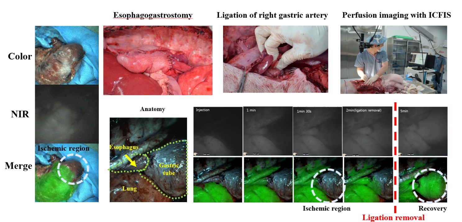
Background : Esophagogastrostomy is the most common esophageal reconstruction procedure after esophagectomy in the early stages of esophageal cancer. After esophagogastrostomy, the insufficient blood supply in the gastric tube may cause anastomotic leaks, a significant complication. It is, therefore, crucial to ensure tissue perfusion intraoperatively for successful anastomosis. We evaluated the feasibility of an intraoperative color and fluorescence imaging system (ICFIS) for evaluating the perfusion of the gastric tube during esophagogastrostomy.
Methods : Ten pigs underwent esophagogastrostomy, and their right gastroepiploic arteries were ligated to mimic ischemic condition of the gastric tube. Indocyanine green (ICG) was used as a near-infrared (NIR) fluorescent tracer and injected intravenously. After 1~5 min, the fluorescence signal-to-background ratios (SBR) both in the gastric tubes and in the esophagus were measured using the ICFIS. In addition, we estimated the time required to acquire the fluorescence signals in 10 pigs that underwent esophagogastrostomy after peripheral (n = 6) or central (n = 4) venous ICG injection.
Results : In all pigs, SBR of the esophagus was much higher than that of the gastric tube, which helped in clearly identifying the ischemic areas in real-time. We observed the recovery of blood perfusion within a few minutes after releasing the ligation of the right gastroepiploic artery. And, time to reach detectable fluorescence signals was shorter in the central route than the peripheral one.
Conclusion : Real-time NIR fluorescent imaging using ICG provides vital information related to ischemia in the gastric tube during esophagogastrostomy. This study provides the ICG dose and preferred injection routes.
책임저자: Hyun Koo Kim
Department of Thoracic and Cardiovascular Surgery, Korea University Guro Hospital, Korea University College of Medicine, Seoul, Korea
발표자: Hyun Koo Kim, E-mail : kimhyunkoo@korea.ac.kr