


초록접수 현황
| 15F-093 | 구연 발표 |
3-Dimensional Image Rendering of Pectus Deformity: A Conceptual Analysis by Combining Morphological Classification and Computerized Tomogram Indices from 4 Different Perspectives
박형주, 김경수, 문영규, 최국빈, 최항준, 안효찬
가톨릭대학교 의과대학 서울성모병원 흉부외과학교실
Background : Morphological evaluation of different types of pectus deformities is crucial for successful repair of pectus excavatum. We developed a morphological classification and computerized tomogram (CT) Indices for better understanding of the malformation, but one of these systems alone only provides a limited information of the real deformity. To elucidate a 3-dimensional image of the deformity, we developed a scheme that combining morphological classification and CT Indices from the author's previous works.
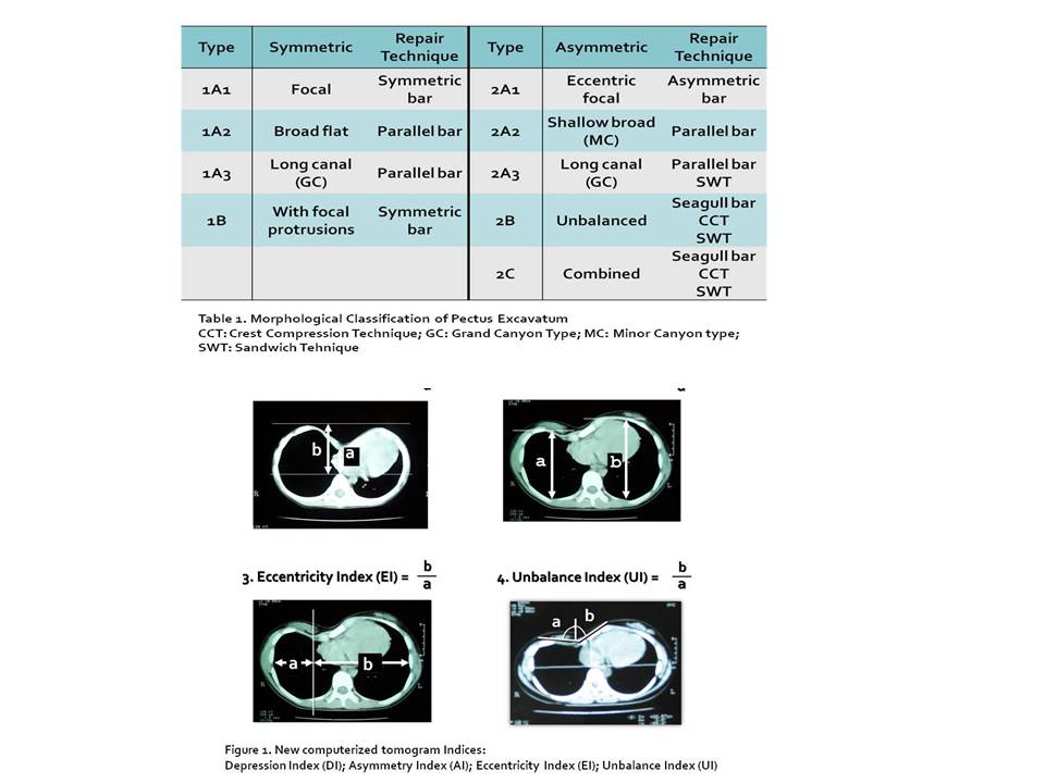
Methods : From our database of pectus excavatum repair, the data of 2419 patients repaired between 1999 and 2014 were retrospectively analyzed. The morphological classification, first established in 2000 and revised in 2012 (Table 1), and 4 CT Indices (Figure 1) were utilized in rendering conceptual 3D images of the deformity.
Results : The mean age of the patients were 9.9 years (range: 16 months to 55 years). Male to female ratio was 3.69. Symmetric type was 1481 (61.2%) and asymmetric type was 938 (38.8%): eccentric 70.0%; unbalance 19.2%; combined 9.5%. Mean values of CTI’s were: DI, 1.84 (1.05~35); AI, 1.05 (1~1.8); EI, 1.54 (1~3.75); UI, 1.50 (1.05~6.0). Morphologic classification provided the basic information whether the deformity was symmetric or asymmetric. In case of asymmetry, it further divided into 3 different types: eccentric; unbalance; and combined. In addition, CTI's were incorporated to render 3D imagination. Depression index furnished how deep the depression was: e.g., DI of 2 represented depressed chest wall is halfway between the sternum and the vertebra. Likewise, asymmetry index, eccentricity index, and unbalance index offered how much each asymmetric component in a quantitative manner for volumetric identification.
Conclusion : With combination of authors' morphologic classification and CTI's rendered 3D image of the deformity and effectively guided selecting appropriate repair techniques for each proposed specific deformity.
책임저자: 박형주
가톨릭대학교 의과대학 서울성모병원 흉부외과학교실
연락처 : 박형주, Tel: 010-4125-2416 , E-mail : hyjpark@catholic.ac.kr
