


초록접수 현황
| 15F-057 | 포스터 발표 |
CT Localization for Patient with Ground Glass Opacity Pulmonary Nodule
Expecting Thoracoscopy: A Mixture of Lipiodol and Indian Ink
김경수¹, 안세하¹, 최항준¹, 백경원², 이교영³, 문석환¹
가톨릭대학교 의과대학 서울성모병원 흉부외과학교실¹, 가톨릭대학교 의과대학 서울성모병원 영상의학과교실², 가톨릭대학교 의과대학 서울성모병원 병리학교실³
Background : Thoracoscopic resections of small or deep seated pulmonary nodules (PNs) sometimes need pre- or intraoperative localization technique to detect and guide the stapling resection. Also, most of PNs can be palpated with finger through the utility incision or thoracoport wound. However, pure ground glass opacity (GGO) lesions are not easy to be palpated and detected by pathologists. We modified the localization technique for detection on surgery and frozen analysis.
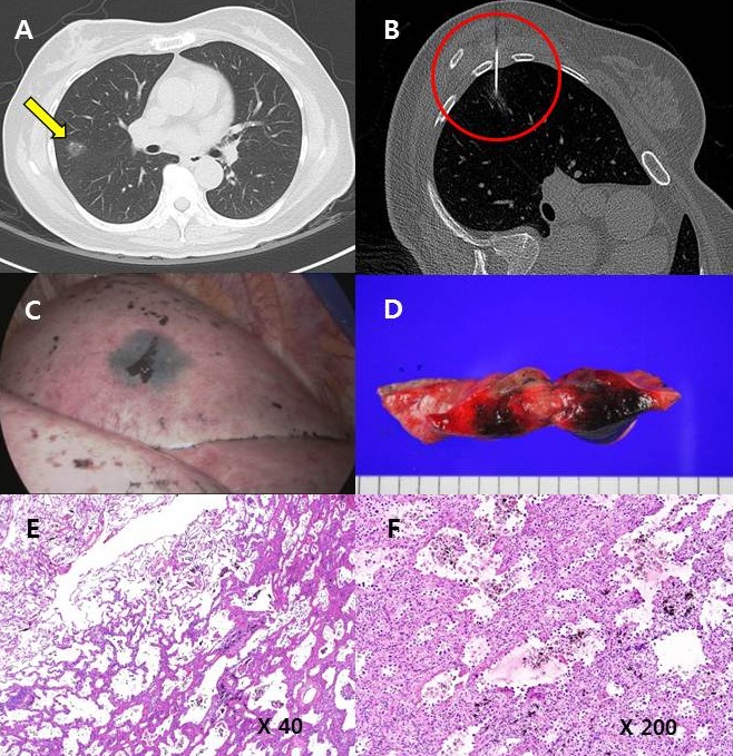
Methods : A 52 year-old female patient presented with a pure GGO lesion which was strongly suggestive of primary lung malignancy on the chest CT scan (Fig. A). We used 0.5 ml of lipiodol and 0.5 ml of indian ink solution. Indian ink solution was prepared in a sterile way how it boiled in a small, stainless steel bowl for heat sterilization for 30 seconds using alcohol. An 18-gauge Chiba needle was percutaneously inserted towards the target using intermittent CT guidance by a radiologist. Once the needle was about 1 mm superior from the target, the mixture solution (0.2 ml) was slowly injected (Fig. B). We intended to mark the visceral pleura near the upper border of the target lesion, rather than the target nodule itself. Another 0.2 ml of the mixture was injected while withdrawing the needle from the lesion, in order to mark the pleura.
Results : This localization procedure took less than 30 minutes, just 30 minutes before transferring the patient to operating room without occurring pneumothorax. Black pigmented tattoo lesion was clearly identified under 2 ports VATS (Fig. C); wedge resection was performed and frozen section revealed a malignancy of adenocarcinoma (Fig. D). Subsequently, VATS lobectomy and mediastinal lymph node dissection was followed. The pathologic result revealed 1.3 x 0.9 cm sized, pT1aN0M0 of stage I, well differentiated adenocarcinoma with lepidic predominant (Fig. E, F). The patient discharged on POD#5 without complication.
Conclusion : We introduced the novel method using a mixture of lipiodol and indian ink solution and we achieved satisfactory result for preoperative localization of GGO PNs prior to VATS surgery. However, further experience will be needed to establish its efficacy and safety.
책임저자: 문석환
가톨릭대학교 의과대학 서울성모병원 흉부외과학교실
연락처 : 김경수, Tel: 02-2258-6139 , E-mail : cskks@catholic.ac.kr
