


초록접수 현황
| 14F-244 | 포스터 발표 |
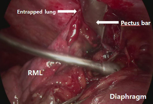
Entrapped Lung between Pecuts Bar and Chest Wall after Pectus Surgery: VATS Findings
김경수, 박형주
가톨릭대학교 의과대학 흉부외과학교실
Background : We incidentally found a partial lung entrapment between the pectus bar and chest wall, or marked adhesions around the pectus bar and adjacent lung in patients who developed pneumothorax after pectus surgery. Through VATS exploration, ruptured bullae seemed to be the cause of pneumothorax, but the role of the entrapped lung as a possible cause of the air leakage was suspected. We report the VATS finding of the lung entrapment.
Methods : (Case 1) An 18 year-old male who had bullae developed pneumothorax on postoperative day 4 after pectus carinatum repair. A VATS exploration on the right lung showed multiple apical bullae with lung entrapment by the pectus bar. The entrapped lung was pulled out and the injured portion removed by wedge resections. One week later, the VATS for the left side pneumothorax identified bullae without lung entrapment.
(Case 2) A 20 year-old male was presented with pneumothorax 2 years after pectus surgery. CT showed no definite bulla but VATS exploration revealed regressed bullae with lung entrapment to the pectus bar. We performed wedge resections for bullae lesion and extracted trapped lung successfully after adhesiolysis.
(Case 3) A 15 year-old male readmitted with bilateral pneumothorax 1 month after pectus surgery. Bilateral VATS exploration showed apical bullae and lung entrapment that multiple wedge resections were followed by adhesiolysis.
Results : Pathologic results identified bullae in all specimens. Postoperative courses were uneventful in all cases.
Conclusion : VATS exploration confirmed lung entrapments by the pectus bar in those patients. These findings demonstrated that pectus repair may complicate with lung entrapment into the pectus bar. However the lung entrapment causes pneumothorax is uncertain, because all the patients had bullae in their lungs.
책임저자: 박형주
가톨릭대학교 의과대학 흉부외과학교실
연락처 : 김경수, Tel: 02-2258-6320 , E-mail : cskks@catholic.ac.kr
